

- 00000018WIA30412970GYZ
- id_400261731.4
- Aug 18, 2022 2:38:08 PM
Magnetization Transfer
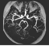
Use the Magnetization Transfer Imaging Option to improve contrast between blood flow and surrounding tissue in 3D TOF images, to augment post-contrast T1-weighted brain images, and to increase myelographic effect for improved disc and cord lesion visualization. This is achieved by saturating tissues containing significant amounts of protein (e.g., gray/white brain matter, skeletal muscle). This is achieved by applying a large saturation pulse that is 1200 Hz off the center frequency.
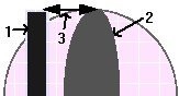
| Number | Description |
|---|---|
| 1 | MT pulse |
| 2 | CSF |
| 3 | pulse offset |
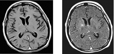
Spin Echo with a Magnetization Transfer preparation technique can be used to augment post-contrast T1-weighted brain images. Similar to the MRA application, the Magnetization Transfer preparation is used to suppress brain parenchyma signal relative to contrast-laden blood.
Magnetization Transfer can also be used with 2D Multi-planar GRE or 3D GRE to increase myelographic effect for improved disc and cord lesion visualization.
- The minimum TR is a function of the gradient strength.
- The off-resonance saturation pulse applies peak SAR. The minimum TR may need to be increased if a FC or spatial saturation pulse is used.
- Magnetization Transfer with 3D TOF GRE/SPGR and 3D GRE/SPGR increases the minimum TR.
- Approximate signal saturation to be expected when using Magnetization Transfer:
- Skeletal muscle: 60%
- White brain matter: 40%
- Gray brain matter: 30%
- Blood: 15%
| PSD | MT Pulse Duration | MT Offset | MT Flip | MR Pulse Type |
|---|---|---|---|---|
| 2D Spin Echo | 16 ms | 1200 Hz | 1100 | Fermi |
| 2D Spin Echo with FC | 16 ms | 1200 Hz | 1100 | Fermi |
| 2D GRE/SPGR | 8 ms | 1200 Hz | 670 | Fermi |
| 3D GRE/SPGR | 8 ms | 1200 Hz | 670 | Fermi |
| 3D TOF GRE/SPGR | 8 ms | 1200 Hz for T/R coils or 2400 Hz for other coils | 950 | Fermi |