

- 00000018WIA30F10970GYZ
- id_400230941.5
- May 4, 2022 1:42:41 AM
Wide bore systems cradle restrictions
The graphic prescription cannot always be acquired due to insufficient cradle scan range. The cradle cannot advance to isocenter if the landmark is less than 23 cm from the edge of the cradle. Below are two examples: feet first head prescription and a head first, lower leg prescription.
Wide bore systems with a stationary table considerations
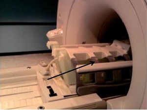
For a lower leg scan with a head first position, there is a landmark limitation tick mark on the table that is 53.3 cm from the foot end of the table.
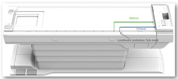
- If the landmark is over the limitation tick mark, then a message may be posted indicating that the scan cradle could not go far enough into the bore to complete the scan.
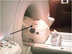
Wide bore systems with a mobile table considerations
For a head scan with a feet first position, the alignment light is positioned at the extreme superior edge of the landmark position, which is approximately 23 cm from the edge of the cradle.
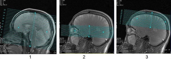
If the graphic Rx slices are positioned at the superior end of the FOV, then a message may be posted indicating that the scan cradle could not go far enough into the bore to complete the scan.
| Number | Description |
|---|---|
| 1 | Entire brain graphic prescription does not present a problem. |
| 2 | Orbit graphic prescription does not present a problem. |
| 3 | Scan prescription centered at the top of the head presents a problem and an error message is posted indicating that the scan table cannot go into the bore far enough to complete the requested prescription. Feet first positioning in this scenario cannot be completed. |
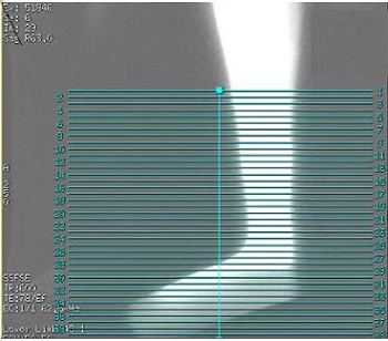
For a lower leg scan with a head first position, the alignment light is positioned at the inferior edge of the landmark position, which is approximately 23 cm from the edge of the cradle.
If the graphic Rx slices are positioned at the inferior end of the FOV, then a message may be posted indicating that the scan cradle could not go far enough into the bore to complete the scan.