- 00000018WIA3096C870GYZ
- id_400229281.4
- Mar 31, 2022 11:30:23 AM
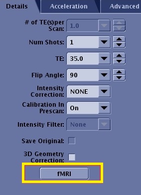
fMRI
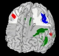
fMRI (commonly referred to as Brain Mapping) is a purchasable option that uses a single-shot SE EPI or GRE EPI pulse sequence in conjunction with neural activation paradigms to visualize signal intensity changes in the brain during task performance. As the patient performs a predefined task, the ratio of oxygenated to deoxygenated hemoglobin varies. The signal changes observed during task activation are due to a complicated interaction of blood flow, blood volume and oxygen uptake.
The fMRI option is used to acquire BOLD images to map task-activated regions in the brain. The BOLD fMRI technique records signal intensities in the brain while the patient is performing a task. The acquired data is processed to yield color maps, which display the levels of neural activation seen in different regions of the brain. This aids physicians in surgical planning, monitoring, and post-surgical monitoring of patients.
The fMRI button on the scan Details tab opens the fMRI Scan Control screen. The fMRI button only appears on the scan parameters Details tab if the protocol has the fMRI Imaging Option selected.