

- 00000018WIA30E66970GYZ
- id_400216641.3
- May 10, 2022 4:18:53 AM
SAT Gap
Spatial saturation (often referred to as SAT) pulses suppress the signal from fat. SAT Gap allows you to adjust the gap between the SAT pulse and the excited slice to maximize Fat SAT effectiveness. By taking advantage of the chemical shift properties inherent to the SAT pulse and its relative position, the fat is suppressed.
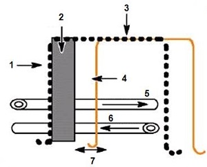
| Number | Description |
|---|---|
| 1 | Fat SAT component |
| 2 | Excited slice |
| 3 | Single SAT pulse |
| 4 | Water SAT component |
| 5 | Desired flow |
| 6 | Undesired flow |
| 7 | 10 mm SAT gap |
- Increase the SAT Gap as the area to be scanned gets farther away from the heart. A 10 mm SAT Gap is typically used for carotid and iliac vessel exams and a 20 mm SAT Gap is typically used for distal femoral and popliteal vessel exams.
- The effectiveness of the fat saturation is maximized at a 10 mm SAT gap. As the SAT Gap increases, the fat suppression becomes less effective.
- In regions of highly pulsatile flow (e.g., popliteal, iliac), a narrow SAT Gap can result in pulsatile artifacts due to saturation of retrograde flow. To reduce the artifact, increase the SAT Gap, which moves the SAT pulse farther away from the slice. As the SAT Gap increases, the ghosting from retrograde flow decreases, but so do the fat suppression effects.