

- 00000018WIA30CFB770GYZ
- id_400218601.4
- Feb 21, 2022 6:31:11 PM
MAGiC warning and considerations
- MAGiC processed images are similar but not identical to images that are acquired with a particular PSD to produce a PD, T1, T2, FLAIR, STIR, PSIR or DIR contrast.
- As is standard clinical practice, it is recommended that all image series generated by MAGiC be evaluated in order to ensure an appropriate level of information is utilized during image review.
- An increased rate of occurrence for MAGiC T1 FLAIR and T2 FLAIR artifacts may be observed as compared to conventional imaging.
- The MAGiC image acquisition for quantification suppresses the signal from moving blood. This results in low signal in vessels with normal blood flow, such as arteries, which will not be enhanced. Usage of MAGiC post injection of a contrast agent has not been evaluated by GE and should not be used.
- Vessels may be suppressed and appear black as an inherent feature of the MAGiC technique.
- MAGiC creates synthetic images that do not contain acquisition protocol information.
- Use ProtoCopy to copy the scan parameters from a MAGiC scan series.
| Warning | |
|---|---|
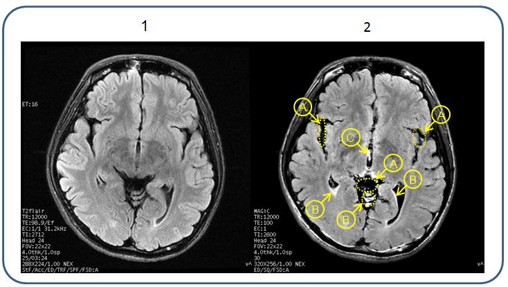
The figure below illustrates an example of differences between a MAGiC T2 FLAIR image compared with a conventional T2 FLAIR.
| Number | Description |
|---|---|
| 1 | Conventional T2 FLAIR |
| 2 | MAGiC T2W FLAIR A = Hyperintense Vessel Sign (HVS) B = High signal intensity at the edge of Cerebral Spinal Fluid (CSF) C = CSF |
Flow artifact considerations
The MAGiC MDME sequence is susceptible to flow artifacts, as is any FSE technique. Due to the application of multiple inversion pulses, this is more apparent in certain situations on MAGiC-derived series
The figure below illustrates an artifact related to pulsatile flow phenomena. Image 1 shows a MAGiC T1 FLAIR image with artifact likely resulting from fast flowing blood. Image 2 shows a MAGiC T2 FLAIR displaying artifact related to CSF flow. Images 3 and 4 present with a notable reduction in artifact as a result of using the recommended parameter settings.
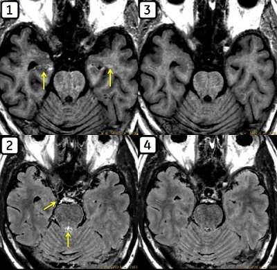
| Number | Description |
|---|---|
| 1 | T1 FLAIR |
| 2 | T2 FLAIR |
| 3 | T1 FLAIR + I SAT |
| 4 | T2 FLAIR + I SAT |
Recommendation: Minimize this artifact by maintaining a slice gap of 10% or larger of the acquired slice thickness. The addition of an inferior saturation region within the imaging FOV can aid in suppressing this artifact. You may also acquire MAGiC in a different orientation for further artifact reduction.
Motion related artifact
Recommendation: Take extra steps to immobilize the patient with extra head padding and if possible, acquire the MAGiC sequence in the beginning of the exam. Acquiring MAGiC in an additional imaging plane or rescanning the series may be needed in the presence of motion. You may also consider using alternate/conventional techniques to resolve such artifacts.
Tissue contrast
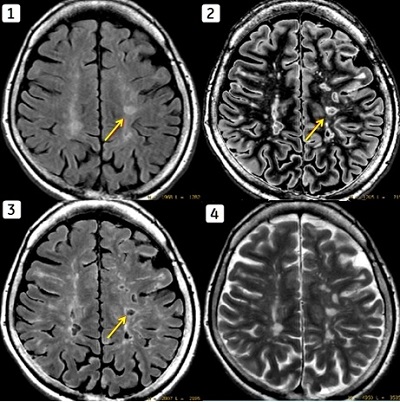
The figure below illustrates an example of a lesion, as visualized on a conventional T2 FLAIR image and multiple calculated MAGiC series. Note the similar performance of DIR to conventional FLAIR.
| Number | Description |
|---|---|
| 1 | Conventional T2 FLAIR |
| 2 | MAGiC DIR |
| 3 | MAGiC T2 FLAIR |
| 4 | MAGiC T2 |