

- 00000018WIA30DCE870GYZ
- id_400224561.5
- May 6, 2022 3:35:08 AM
SAT: chemical
Chemical saturation takes advantage of the fact that fat and water precess at different frequencies.

| Number | Description |
|---|---|
| 1 | Water peak |
| 2 | Fat peak |
| 3 | Water/fat separation: 220 Hz at 1.5T |
The success of spectral saturation techniques depends on the uniformity of the anatomical area being imaged, in addition to the pulse sequence and coil being used. While the system is shimmed to a system specification to provide you with optimal homogeneity, once a patient is placed in the magnet bore, the homogeneity can be affected. For example, an abdomen may be more uniform than a shoulder. It works best with anatomy of interest at isocenter and a small FOV.
Fat or water suppression reduces chemical shift artifacts because these artifacts are caused by relative shifts of fat from water. When one component is suppressed, there is nothing for the other component to shift away from.
Chemical suppression decreases slice locations per TR for two reasons: the extra time required to apply the RF pulse and chemical suppression pulse's contribution to Specific Absorption Rate (SAR).
Site or patient specific inhomogeneities may be unavoidable even at isocenter. The result can be uneven suppression.
There are several types of Chemical SAT:
- Fat
- Fat Classic
- Water
- SPECIAL
Fat/Water considerations
- Fat and water cannot be selected at the same time.
- Fat/water suppression selections turn off when the PSD changes.
- For T2-weighted scans, it is advisable to lower the TE to approximately 75 ms. The T2 of muscle is such that it loses signal at longer TEs.
- Use Echo Train Lengths (ETLs) of 8 to 12. Long ETLs average in signal from very late echoes, resulting in decreased signal from non-fatty tissue.
- The overall Signal-to-Noise Ratio (SNR) of improved Fat SAT images may slightly decrease. Consider adjusting parameters which affect SNR, e.g., increase NEX, increase the FOV, decrease matrix size, decrease the Receive Bandwidth.
- The Fat SAT improvements decrease the possibility of poor or uneven saturation; however, tissue saturation may be reduced when the FOV is greater than 20 cm.
- Uneven saturation can still occur as a result of local inhomogeneities, e.g., at air/tissue interfaces or when the anatomy of interest is non-uniform.
- For FSE T2 fat suppressed accusations with Improved Fat SAT, non-fatty tissue (muscle in particular) may appear darker than desired.
SPECIAL considerations
- There is a slight increase in scan time to account for the inversion pulse that is applied once every 64 slice encodings. This increase in scan time is much shorter than the increase that would occur if a FAT SAT pulse was used.
- SPECIAL supports manual tuning for center frequency adjustment but does not support manual tuning for flip angle adjustment.
- SPECIAL is not available with all pulse sequences.
ASPIR considerations
Consider the following with SPECIAL and 2DFSE-ASPIR:
- Adiabatic Spectral Inversion Recovery (ASPIR) use a spectral-selective adiabatic RF pulse to invert only fat signal thus producing a more uniform fat suppression in comparison to non-ASPIR techniques. The image acquisition is turned on at the inversion time when the fat signal is at the null point.
- There is an approximately 30% increase in scan time to account for the ASPIR inversion pulse.
- ASPIR is more robust for B1 inhomogeneities when compared to Chemical Fat Saturation (CFS). ASPIR is particularly appealing when acquiring scans in anatomical areas such as the pelvis where B1 inhomogeneities often cause non-uniform fat suppression with a CFS technique. The pelvis images compare the results of Chemical Fat Saturation and ASPIR. Note the apparent areas of non-uniform Fat Sat suppression in the CFS image. The ASPIR image has improved fat suppression, but the scan time increased approximately 30%.
Figure 2. Chemical fat saturation compared to ASPIR in the pelvis 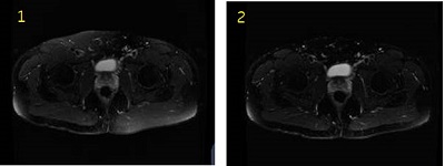
Table 2. Image legend Number Description 1 Image acquired with CSF technique. Note: the very apparent areas of non-uniform fat suppression.2 Image acquired with ASPIR technique. Note: the uniform fast suppression. The scan time was increased approximately by 30%. - 2D FSE ASPIR is only available with T2-weighted and PD-weighted 2D FSE protocols that have a TR greater than 1500 ms and an ETL greater than 5.
- Fat SAT Efficiency can be used to modify the amount of fat saturation. As the value increases, fat becomes darker on the image. As the fat saturation value increases, more fat is suppressed.
- SPECIAL when used with VIBRANT type-in PSD Name efgre3d_aspir, COSMIC, Inhance 3D Inflow IR, SSFSE, 2D FIESTA on 3.0T systems, and Diffusion Weighted Imaging uses an ASPIR fat suppression technique.
- Cube used with Fat SAT or Fat Classic uses an ASPIR fat suppression technique.