

- 00000018WIA3041D770GYZ
- id_400247141.3
- Apr 5, 2022 11:21:16 AM
Mode
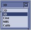
The Imaging Mode defines the image format and type of image information to be gathered. The Mode text box has predefined values to select 2D, 3D, Cine, Calib, or MR Spectroscopy.
2D
2D mode acquires and reconstructs raw image data into two-dimensional images, whose brightness is proportional to the intensity of the MRI signal from the corresponding protons. The RF pulse and gradient pulse occur at the same time to excite an individual slice of a specific thickness of tissue.
3D
3D mode excites an entire scan volume or slab with an RF pulse and spatial encoding is performed in the phase, frequency, and slice axes.
MRS
MRS (Magnetic Resonance Spectroscopy) mode is an optional software package. It acquires a volume-localized, water-suppressed spectrum from a single or multiple voxels or a volume of interest.
Calibration
Calib (Calibration) mode automatically completes scan parameter fields other than slice thickness and start and end locations for a calibration scan.
Cine
Cine mode is an optional software package that lets you generate images for dynamic views of anatomy such as the heart. This option employs retrospective gating techniques. It is compatible with Vascular and Gradient Echo family pulse sequences.
Cine mode is only compatible with GRE pulse sequences that acquire data continuously throughout the cardiac cycle. Cine scans employ a short TR GRE pulse sequence that produces a bright blood and dark myocardium image with fairly high contrast. Cine images are acquired using retrospective gating techniques.
Cine mode acquisitions are neither gated nor triggered. When the Cine imaging mode is selected, the cardiac cycle is monitored and data acquisition takes place throughout the cycle. Data is collected continuously throughout the cardiac cycle and is retrospectively sorted to place the acquired data synchronous to the averaged cardiac cycle. An Arrhythmia Rejection Window (ARW) is defined at the Combined Card/Resp screen. If an R-wave occurs outside the ARW, the image data collected during that period is discarded. This data must be collected again, resulting in extended scan time. See Cine tab.
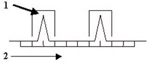
| Number | Description |
|---|---|
| 1 | Arrhythmia Rejection Window |
| 2 | TR |
To view the Cine mode tab, from the Scan Parameters screen menu bar, click the  and then click the Cine tab. The Cine tab only appears if the protocol has the Cine Imaging Mode selected with a GRE, SPGR, or Phase Contrast pulse sequence.
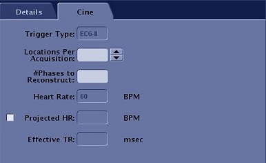
| Parameter | Description |
|---|---|
| Trigger Type | Displays the gating sensor selected from the Waveform and Gating Selection area of the Gating Control screen. This field cannot be edited or stored in a protocol. Make your gating selection from the Gating Control screen during patient setup. That selection remains active until it is changed. |
| Locations Per Acquisition | In the Locations Per Acquisition text box, type in the number of slice locations to acquire in each acquisition. The minimum and maximum values are displayed next to the text box. The number of locations per acquisition affects scan time, the Effective TR, and contrast between blood and muscle tissue. As the number of locations per acquisition increases, the Effective TR increases and blood/tissue contrast decreases. Consider two slice locations per acquisition to produce images exhibiting the best compromise between image quality. |
| # Phases to Reconstruct | In the # Phases to Reconstruct text box, enter the number of cardiac phases to obtain; up to 32 phases per cardiac cycle for each slice. Image quality may decrease as the number of cardiac reconstructed phases increases. Sixteen is a typical number and a compromise between image quality and the need for an adequate number of phases. |
| Heart Rate | Displays the patient's heart rate. The heart rate is automatically updated at 5 second intervals only if the Cardiac tab is open. This is not an editable field. If you copy/paste a series from the Workflow Manager, the heart rate is copy and pasted along with all other scan parameters. Therefore, if you anticipate the heart rate is going to change, open the Cardiac tab so that the Heart Rate field updates every 5 seconds. |
| Projected HR | Type in a projected heart rate in the text box, if necessary for the exam. For example, stress exams or when you want to scan with a specific heart rate due to a significantly fluctuating heart rate. |