

- 00000018WIA30FFD870GYZ
- id_400221371.5
- May 25, 2022 4:00:23 AM
FSE Double and Triple IR scan
FSE Double and Triple IR use a non-slice-selective IR pulse to invert all spins in the body, including blood. A second slice-selective IR pulse is immediately applied, re-inverting spins in the image slice. At this point, magnetization within the slice is essentially unchanged, as compared to the state of the spins prior to the initial whole body IR pulse.
A BSPTI delay time is set to equal the time required to allow inverted blood spins to reach the null point, about 650 ms with a 60 BPM heart rate. The BSP TI time occurs during the systolic portion of the cardiac cycle, resulting in a “wash-in” and “wash-out” of blood; the nulled blood flows into the imaging slice, while the blood in the slice during the slice re-inversion IR pulse exits. After the BSP TI time, the FSE sequence is initiated and image data is acquired as in any FSE acquisition.
Triple IR uses a third IR pulse (TI Time field) to null fat signal in the slice.
Use Double and Triple IR scans to visualize cardiac anatomy, myocardial wall masses, valve leaflets, and black blood. FSE Double and Triple IR images are very sharp images in comparison to SSFSE Double and Triple IR scans, but they are longer acquisitions.
| Number | Description |
|---|---|
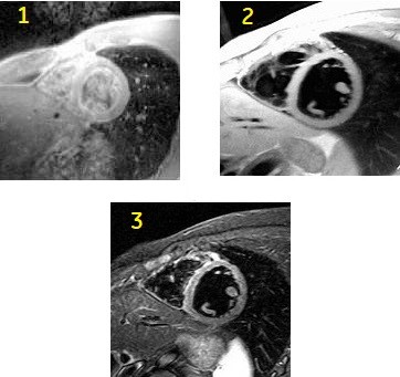
| 1 | FSE short axis cardiac image |
| 2 | Double IR short axis cardiac image |
| 3 | Triple IR short axis cardiac image. |
Consider this information when modifying FSE Double Triple IR scan parameters. For specific scan parameter values, select a protocol from your GE or Site library.
- Scan selections for Double-IR: 2D Mode, Fast Spin Echo family, FSE pulse, Blood Suppression Imaging Option.
- Scan selections for Triple-IR: 2D Mode, Fast Spin Echo family, FSE pulse, Blood Suppression and IR Prepared Imaging Option.
General considerations
- One slice is acquired per acquisition, therefore cross-talk is not an issue.
- If more than one location is prescribed (as is generally the case), select a number of locations before pause to allow for breath hold instructions.
Scan parameters
- Bandwidth: As receive bandwidth increases, the ESP decreases (which is desirable), and the maximum BSP TI increases. Blood Suppression acquisitions generally use maximum bandwidths to keep the echo space small and thereby decrease the effects of blurring with the long ETLs. Compensate for the loss in SNR that occurs with these wide bandwidths by increasing the slice thickness or FOV.
- Fat/Water SAT: Chemical SAT pulses (Fat/Water Suppression) are not available for FSE-IR with Blood Suppression (Triple-IR Blood Suppression).
- Fat/Water SAT: Chemical SAT can be used for FSE with Blood Suppression (Double-IR Blood Suppression).
- Phase FOV: When using Torso Phased Array Coil, use a 1 Phase FOV to avoid wrap-around artifact.
- TE: TE values of 40 ms or greater are likely to reduce the appearance of flow related artifacts.
- TI: Triple IR: the Inversion Time for nulling fat at 3.0T is approximately 230 ms. This is the same TI time used in other short TI inversion recovery sequences when fat nulling is desired.
- TI: The BSPTI Auto selection prompts the system to calculate the most accurate value based on the patient’s heart rate.
- A time course study decreases the T1 of blood, which may require a decreased BSP TI. The Auto calculation assumes that the series is not a time course study. The Auto BSP TI selection is calculated to obtain maximum blood suppression. If the calculated value is too high for selected scan parameters, then the BSP TI must be decreased by increasing the bandwidth, decreasing the ETL, and/or the Trigger Window.
- It may be necessary to increase the RBw or decrease the ETL to obtain the proper BSP TI when the heart rate is > 100 BPM and the TW is wide. A message is posted when this is necessary.
Gating tab
- Trigger Delay: Diastolic and Systolic Trigger Delays are available. For optimum image quality and very good blood suppression in the ventricles, select Trigger Delay: Diastolic.
- RR-interval: Using a 1 RR interval allows for shorter scan times and therefore it is useful for breath hold scans. A single RR provides a more T1-weighted appearance. However, images are acquired at early rather than late diastole which may degrade image quality. Typically, only use 1 RR black blood technique for applications where it is necessary to acquire a short breath hold scan.