

- 00000018WIA30446970GYZ
- id_400236181.3
- Aug 21, 2022 5:40:51 PM
Vascular
The Vascular family of pulse sequences can be used during MRA imaging for optimal visualization of the vascular structures of interest. The Vascular pulse sequences can be acquired in the 2D, 3D, and Cine imaging modes.
| PSD | Anatomic region | Acquisition |
|---|---|---|
| 2D TOF | Carotid bifurcation, venous anatomy, aortic arch, peripheral vessels | Multiple thin slices displayed as maximum pixel projections of the volume imaged |
| 3D TOF | Circle of Willis, abdominal vasculature | Volume SPGR or GRE data set displayed as maximum pixel projections |
| Multi-slab TOF | Intracranial vasculature, carotid bifurcation, aortic arch, peripheral vessels, venous anatomy | Multiple overlapping axial GRE volumes displayed as maximum pixel projections |
| 2D Phase Contrast | Localizer/flow direction and velocity for intracranial and extracranial vasculature, portal or hepatic vein, quantitative measurement of flow velocity | Multi-slice or thick slab projection image |
| 3D Phase Contrast | Intracranial vasculature, renal arteries | Volume acquisition obtained with flow encoding displayed as multiple maximum pixel projections |
| Cardiac-gated (cine) 2D Phase Contrast | Aortic arch, peripheral vessels, quantitative measurement of flow velocity over cardiac cycle | PC images obtained at different cardiac phases |
Blood flow terms
- Viscosity
- The resistance of blood flow due to the friction of blood elements in a moving stream. Blood viscosity decreases in cases of anemia and increases in conditions such as polycythemia. Turbulent blood flow is more frequently encountered in low-viscosity conditions.
- Laminar Blood Flow
- The distribution of flow velocities along the vessel layers. Velocities are slowest along the vessel wall and most rapid within the central portions of the vessel.
- Peak Velocity
- The maximum velocity encountered within the lumen of the vessel under consideration. Peak flow velocities vary with exercise, anatomic location, and pathological conditions. The ascending aorta has the highest velocities. Generally, as you move distally from the heart, the number of vessels and total area increase, decreasing flow velocities.
- Turbulence
- Chaotic flow with randomly fluctuating velocity components. At normal blood flow velocities, laminar flow predominates, then as velocity increases and exceeds a critical threshold, turbulence is encountered. Turbulence can compromise MRAs.
Figure 1. Turbulent flow occurs distal to areas of stenosis. Vortex flow is created as blood suddenly decelerates in areas of post-stenotic dilatation 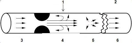
Table 2. Image legend Number Description 1 Vortex flow 2 Distal to stenosis 3 Laminar 4 High velocity 5 Turbulent 6 Laminar
Flow patterns
Complex flow may cause areas of reduced signal intensity within the vessel lumen in MRA imaging, therefore, it is important to consider vessel flow patterns.
- Vortex Flow
- Localized, slowly swirling or stagnant blood flow that occurs distal to areas of arterial stenosis and at sites of arterial bifurcations.
- Flow Separation
- Streamline flow separates from the wall of the vessel creating a separated region of complex flow with eddying motion, counter-current flow, and reduced velocity.