

- 00000018WIA30393970GYZ
- id_400226441.4
- Mar 2, 2022 12:20:46 PM
SSFSE and SSFSE-IR black blood scan
Use SSFSE and SSFSE-IR for black blood applications: heart and vascular structures. Black blood MRI scans are used to null the signal of flowing blood. In addition to suppressing signal from flowing blood, SSFSE-IR also suppresses fat signal. SSFSE with Blood Suppression is a faster scan in comparison to FSE. The SSFSE scan can be acquired within a breath hold or even as a free-breathing scan.
Black blood double-IR
- Black blood double-IR acquires 8-10 slices in a single breath hold (15-20 seconds).
- Scan selections: 2D Mode, Fast Spin Echo family, SSFSE pulse, Imaging Options: Blood Suppression (Cardiac Gating and Sequential are on and cannot be edited).
- Diastolic and Systolic Trigger Delays are available. For optimum image quality and very good blood suppression in the ventricles, select Trigger Delay: Diastolic.
Figure 1. Image acquired with Diastolic Trigger Delay 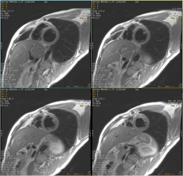
- SSFSE can be acquired with a fat suppression technique: Fat SAT, SPECIAL or change the PSD to SSFSE-IR.
Figure 2. Good fat and blood suppression with SSFSE and fat saturation techniques 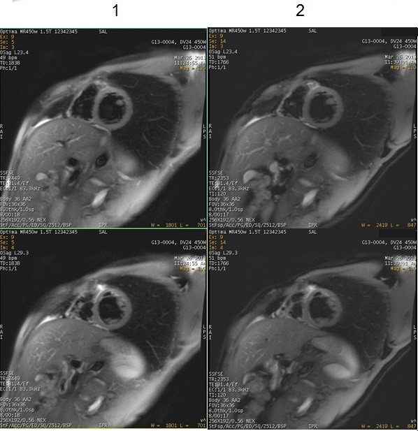
Table 1. Image legend Number Description 1 SSFSE with Blood Suppression Imaging Options and Fat SAT. 2 SSFSE with Blood Suppression Imaging Options and Chemical saturation SPECIAL. SPECIAL is the preferred fat saturation technique because it uses an ASPIR pulse, which is insensitive to B1 inhomogeneities. For more details about ASPIR, see ASPIR considerations.
- Diastolic and Systolic Trigger Delays are available. For optimum image quality and very good blood suppression in the ventricles, select Trigger Delay: Diastolic.
Black blood triple-IR
- Black blood triple-IR acquires 8-10 slices in a single breath hold (15-20 seconds).
- Scan selections: 2D Mode, Fast Spin Echo family, SSFSE pulse, Imaging Options: IR-Prepared, and Blood Suppression (Cardiac Gating and Sequential are on and cannot be edited).
- Use SSFSE-IR for fat suppression when B0 inhomogeneity is a concern.
- An image degradation artifact is possible if the Inversion Delay for blood suppression is not appropriate or the Inversion Time for fat suppression is not proper.
Figure 3. SSFSE-IR scans acquired with varying TI times 
Table 2. Image legend Number Description 1,2,3 Note: the out-of-phase artifact seen when the TI is set to 120 ms, 140 ms and 150 ms.4 Optimum image quality when the TI is set to 130 ms.
- ARC and ASSET are available if a compatible coil is selected.
- The BSPTI Auto selection prompts the system to calculate the most accurate value based on the patient’s heart rate.
- A time course study decreases the T1 of blood, which may require a decreased BSP TI. The Auto calculation assumes that the series is not a time course study. The Auto BSP TI selection is calculated to obtain maximum blood suppression. If the calculated value is too high for selected scan parameters, then the BSP TI must be decreased by increasing the bandwidth, decreasing the ETL, and/or the Trigger Window.
- It may be necessary to increase the RBw or decrease the ETL to obtain the proper BSP TI when the heart rate is > 100 BPM and the TW is wide. A message is posted when this is necessary.
User CVs
Click the Advance tab to view the available User CVs. The CVs may vary based on the field strength and selected scan and imaging parameters.
The User CVs listed below are available with Blood Suppression Imaging Option selected.
- Collect All Available Echoes
- Invert Slice-selection Gradients
- Maximum Number of Echoes
- PURE compensation
- Slice Optimization
- Use when Tailored RF Imaging Option is turned off.