

- 00000018WIA308F3970GYZ
- id_400243471.6
- Aug 20, 2022 11:13:43 AM
SPGR scan
SPGR is a GRE sequence that uses continuous phase shifting of the RF excitation pulse to spoil residual transverse magnetization. The RF spoiler pulse, minimum TE, short TR (40 to 60 ms), and moderate flip angle (30 to 50°) result in T1-weighted image contrast. SPGR is available in 2D sequential and 3D modes.
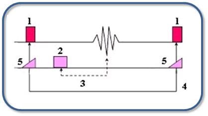
| Number | Description |
|---|---|
| 1 | Variable pulse |
| 2 | Gradient pulse |
| 3 | TE |
| 4 | TR |
| 5 | Phase shift |
SPGR is used to acquire T1-weighted contrast images. Sequential SPGR acquisitions eliminate cross-talk because all data is obtained one slice at a time.
Consider this information when modifying SPGR scan parameters.
- Scan selections: 2D or 3D Mode, Gradient Echo family, SPGR pulse.
General considerations
- As flip angle decreases, SNR decreases. SPGR signal reduction requires a surface or extremity coil, more NEX, or 3D mode.
Figure 3. TR/T1 and Flip Angle Curve 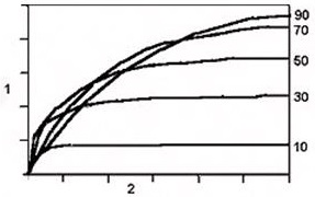
Table 2. TR/T1 and Flip Angle Curve image legend Number 1 Signal in 0.2 increments 2 TR in 500 ms increments
Scan parameters
- Flip angle: It affects the amount of recovery that occurs between each excitation pulse. As a general rule, the higher the flip angle the more saturation and T1 effects are seen in the image.
- TR and Flip angle: 2D sequential or 3D mode: keep the TR and flip angle within 10 points of one another to produce the optimum SNR.
- TE: SPGR scans are more sensitive to any process that causes signal loss due to B0 inhomogeneities, chemical shift, and magnetic susceptibility effects. These effects increase as TE increases. Signal voids are seen particularly where there is metal in the body and air/tissue interfaces.
Imaging Options
- Motion Compensation: Select it to reduce respiratory motion and residual cardiac motion. For details, see Motion Compensation.
2D SPGR User CVs
Click the Advance tab to view the available User CVs. The CVs may vary based on the field strength and selected scan and imaging parameters.
- When Magnetization Transfer Imaging Option is turned On, then these CVs are available:
3D SPGR User CVs
Click the Advance tab to view the available User CVs. The CVs may vary based on the field strength and selected scan and imaging parameters.
- Apodization Level
- Image acquisition delay
- Maximum Monitor Period
- Use with SmartPrep
- Real-time SAT
- Restricted Real-time Navigation
- Slice Partial Fourier
- Turbo Mode
User CVs with Respiratory and Cardiac Gating Imaging Options selected
User CVs with Navigator Imaging Options selected