

- 00000018WIA3080E870GYZ
- id_400249081.2
- Mar 2, 2022 9:52:00 AM
2D FIESTA scan
The FIESTA sequence is a fully balanced steady-state coherent imaging pulse sequence designed to produce high SNR images at very short TRs. It is a fast-ECG-gated, segmented k-space acquisition. It accentuates the contrast of spins with high T2/T1 ratios (such as cerebral-spinal fluid, water, and fat) while suppressing signal from tissues with low T2/T1 ratios (such as muscle and myocardium).
Use 2D FIESTA for imaging abdomen and cardiac function in which clear delineation between the blood (bright) and the myocardium (dark) is needed. It is also useful for valve assessment cardiac imaging, as FIESTA compensates for turbulent flow. Water and fat contrast is accentuated, while muscle and myocardial tissues are suppressed.
Consider this information when modifying 2D FIESTA scan parameters. For specific scan parameter values, select a protocol from your GE or Site library.
General considerations
- Scan selections: 2D Mode, Gradient Echo family, Fiesta pulse.
- Bandwidth: The default is 125 kHz, range = 83.3 to 125 kHz, depending on system configuration. Reducing the bandwidth increases the TR and slightly increases SNR.
- Flip angle: When the flip angle ≥ 50°, the SAR is affected resulting in a longer TR.
- Phase/Frequency: The Phase can be > Frequency value.
- Lower frequency values allow a shorter TR and therefore a shorter scan time. This in turn can minimize flow artifact.
- Slices: Slices are acquired sequentially and cross-talk is not a problem. As FOV decreases, TR increases.
- Sequential scanning acquires one slice per acquisition. The number of slices indicates the number of acquisitions prescribed.
- After you prescribe the slices, return to Locs before Pause and prescribe a pause in the scan at predetermined points for breath hold studies.
- TR: The advantages of FIESTA can only be realized with a very short TR. The minimum TR is selected automatically. The calculated TR is the minimum TR attainable and is based on SAR restrictions. The TR may be adjusted through changes to any of the following parameters: frequency matrix, FOV, slice thickness, and flip angle. You achieve the best image quality at TR ≤ 3 ms at 3.0T. The bandwidth is typically ≥ 125 to keep the TR < in the recommended range.
Cardiac
- Motion Compensation: Select it to reduce respiratory motion and residual cardiac motion. For details, see Motion Compensation.
- TE: Selecting TE = Minimum may achieve shorter TRs.
- Phase: As phase increases, scan time increases, which can be compensated for by increasing the VPS.
- Views per Second: Recommendations:
- BPM ≤ 60, use 16-26 VPS
- BPM = 61-94, use 16-24 VPS
- BPM > 95, use 14-20 VPS
Abdominal
- ASSET: Select the Imaging Option if desired and if your coil is ASSET compatible.
- SAT: De-select Cardiac Gating to activate SPECIAL in Graphic Rx.
- Slice spacing: Zero or a negative spacing is allowed.
- Slice thickness: As slice thickness decreases, TR may increase. To keep the TR as low as possible, consider increasing BW, decreasing frequency matrix, increasing FOV.
- Slices: When single slice with MPH is selected or multi-slice with MPH+Sequential mode is selected, the chemical saturation effects result is different between the first phase and the other phases. This is due to the saturation of the fat signal. It is recommended that you prescribe at least 2 slices with interleave acquisition for MPH scans.
Figure 2. 2D Fat SAT Fiesta with MPH in sequential mode 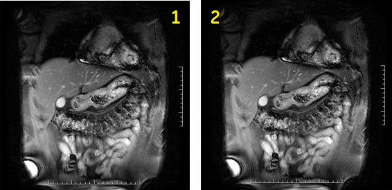
Table 1. Image legend Number Description 1 First phase 2 Second phase Figure 3. 2D Fat SAT Fiesta with 2 slices and MPH in interleave mode. 1 = 1st phase, 2 = 2nd phase 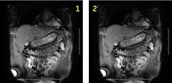
Table 2. Image legend Number Description 1 First phase 2 Second phase - Shim volume: 2D Fat SAT FIESTA is particularly sensitive to shim variations. Therefore it is always recommended to place a shim volume during the graphic prescription process.
Figure 4. Abdominal image comparison 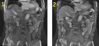
Table 3. Image legend Number Description 1 No shimming prior to the acquisition. 2 Shimming prior to scanning. Notice the absence of the band-like artifact on the image.
Prescan
- 2D Gated FIESTA sequences that are prescribed in a single slice group have a unique prescan and shimming technique. Cardiac FIESTA imaging often results in off-resonance artifacts when the center frequency is incorrectly obtained. These artifacts appear as inhomogeneous areas within the blood pool and as ghosting across the image. They are more prominent in areas of fast or turbulent blood flow.
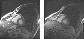
Figure 5. Off-resonance effects on cardiac FIESTA 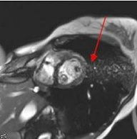
- The enhanced FIESTA sequence significantly reduces off-resonance effects that are sometimes encountered when the correct center frequency of a cardiac image is difficult to obtain. The following enhancements in the 2D FIESTA prescan technique result in a significantly improved spectrum:
- Suppression of fat signal
- Only displaying the frequency spectrum of the relevant FOV
Figure 6. 2D FIESTA: Center Frequency Spectrum 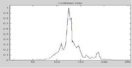
Figure 7. Enhanced FIESTA: Center Frequency Spectrum 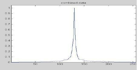
User CVs
Click the Advance tab to view the available User CVs. The CVs may vary based on the field strength and selected scan and imaging parameters.
- Apodization Level
- Use with FIESTA Cine.
- Arrhythmia Monitoring
- Use with FIESTA and cardiac gating.
- Use it with patients that have an irregular heartbeat.
- PURE compensation