

- 00000018WIA30BF3970GYZ
- id_400270131.4
- Aug 20, 2022 11:57:03 AM
Fast SPGR scan
Fast SPGR uses variable flip angles to excite protons, then rephases them by means of gradients. Compared to the SPGR sequence, the Fast SPGR sequence uses a shorter duration excitation RF pulse and a wider receive bandwidth that shortens the duration of the readout time. This implementation allows the prescription of shorter TRs and TEs and therefore shorter total scan times.
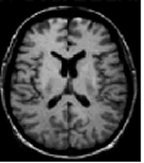
Fast SPGR sequences are used to produce T1-weighting in images where tissues with short T1 are bright and tissues with long T1 are dark. In the brain, white matter is brighter than gray matter and CSF is dark.
Consider this information when modifying Fast SPGR scan parameters. For specific scan parameter values, select a protocol from your GE or Site library.
- Scan selections: 2D or 3D Mode, Gradient Echo family, Fast SPGR pulse.
General considerations
- Fast SPGR sequences result in reduced SNRwhen compared to non-fast SPGR sequences. The SNR decrease results from the use of: higher bandwidths, ultra-short TR values, fractional NEX, and fractional echo.
- Chemical shift effects are seen when a voxel contains both fat and water and the TE is timed for the vectors to be in or out of phase. Boundary between fat and tissues with much water are either bright or dark.
- 3D FSPGR: Type-in PSD 3dradial is used with User CV Chemical Shift Reduction to minimize chemical shift ghosting artifact for fat and water mixed anatomies such as the neck and extremities. For details, see Chemical Shift Reduction.
Scan parameters
- NEX: Increasing NEX to improve SNR may not be an option because of the increased scan time. However, the multi-planar option can be used to improve SNR.
- TR: Due to the short TRs, saturation effects occur resulting in a reduction in SNR and CNR. Short TRs do not allow flip angle flexibility to manipulate image contrast because increasing the flip angle can produce greater saturation effects.
- Bandwidth for 3D FSPGR : As the bandwidth decreases, the following occurs: SNR increases, chemical shift artifact increases, minimum TE increases, which can potentially decrease the number of slices and increase motion artifact. Generally, wider bandwidths are used with Fast sequences to keep minimum TEs and TRs.
- TE for 3D FSPGR: dual echo scans, when TE range reaches it’s in/out of phase limits some imaging parameters are restricted.
- 2 TEs per Scan automatically acquire 1 echo with fat and water out-of-phase and 1 echo with fat and water in-phase.
- Short TEs increase T1 contrast and increase SNR. Increase the TE to increase T2* contrast, decrease SNR, decrease signal changes at fat/water interfaces, and increase magnetic susceptibility effects.
- TR for 3D FSPGR: Short TRs decrease SNR, increase T1 contrast, and decrease scan time. Long TRs increase SNR and scan time.
- TR is not selectable with Tissue Prepared, Multi-Phase, and 3D sequences. The minimum value is set by the system.
- For multi-planar sequences the use of longer TRs (60-100 ms) allows larger flip angles (40-60°), which can improve SNR.
- If the TR and flip angle are within 10 points of one another, the SNR is optimized.
- Prep Time for 3D FSPGR : It only appears if SPECIAL, DE Prepared, or IR Prepared are selected.
- For SPECIAL, select Auto or a T1 in the 30 to 60 ms range, and the system determines the optimum flip angle for the Inversion pulse.
- For IR-Prepared Imaging Option without Cardiac Gating or 3D mode, the Prep Time is calculated from the Inversion pulse to the acquisition of the center of k-space, which is where the contrast is determined. This calculation method is designed to optimize liver/spleen contrast. Typically select a TI value of 500 to 600.
- For DE Prepared Imaging Option, the system sets the time between the first and third prep pulses. Tissue contrast varies as Inversion Time varies.
Imaging Options
- Respiratory Trigger: When it is on, the available imaging time is segmented by the Min TR. It is used to acquire as many phase and slice encodings as possible that will fit in the available imaging time for one respiratory interval.
- Because the 3D dataset is acquired over multiple respiratory intervals, it is recommended that a larger Trigger Window (60%) be used to acquire as much data as possible between respirations.
User CVs
Click the Advance tab to view the available User CVs. The CVs may vary based on the field strength and selected scan and imaging parameters.
User CVs for 2D Fast SPGR
- Apodization Level
- Arrhythmia Rejection
- Use with 3D mode and Navigator Imaging Option selected
- Image Acquisition Delay
- Maximum Monitor Period
- Use with SmartPrep Imaging Option selected
- PURE Compensation
- Real Time SAT
- Use with Fluoro Trigger Imaging Option selected
- Restricted Real Time Navigation
- Use with Fluoro Trigger Imaging Option selected
- Slice Partial Fourier
- Turbo mode
- Chemical Shift Reduction
- Use with type-in psd 3dradial
Post-process tasks
There are multiple compatible post-process tasks. For details, see Add post-process task.
- Maximum Intensity Projection
- Multi Planar Reconstruction
- Image Enhancement Filters