

- 00000018WIA30156970GYZ
- id_400252671.1
- Feb 24, 2022 3:23:54 PM
Fast Card GRE/SPGR scan
Fast Card is a fast, 2D, GRE or SPGR sequence that acquires multiple phases of the cardiac cycle at single or multiple locations. Fast Card segments the number of views into groups of views per segment, with each group of views acquired after a cardiac trigger. Views per segment are the number of k-space lines acquired per cardiac phase, during one RR interval. The Views per Segment parameter affects the temporal resolution, blurring, and scan time of a fast cardiac scan.
Fast Card uses uniform TR to eliminate the lightning-flash artifact that makes the first cardiac phase appear brighter than subsequent cardiac phases.
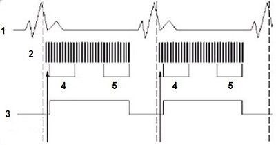
| Number | Description |
|---|---|
| 1 | ECG |
| 2 | RF |
| 3 | Data acquisition |
| 4 | Phase 1 |
| 5 | Phase n |
Use Fast Card for:
- breath-hold cardiac imaging (Fast Card GRE makes blood brighter and SPGR makes the myocardium brighter)
- removing motion in pediatric studies by using Fast Card with multiple NEX
- coronary artery imaging when used with Fat SAT
- cross-sectional studies of the cardiac chambers or the aortic arch
- evaluating cardiac function and valve assessment
Consider this information when modifying FastCard GRE/SPGR scan parameters. For specific scan parameter values, select a protocol from your GE or Site library.
- Scan selections: 2D mode, Vascular family, Fast Card GRE or Fast Card SPGR pulse.
General
- Unless the heart rate varies and causes a trigger outside of the Trigger Window, the RF signal is not maintained through the QRS complex.
- The system pauses when the expected signal is not detected.
- If more than 4 arrhythmias occur for scan times < 25 seconds, the scan aborts.
- Sequential sequences decrease TOF effects for slow or in-plane flow and produce poor flow contrast in longitudinal plane.
- Sequential Fast Card is typically used to acquire short axis and non-sequential is used to acquire a localizer.
- Non-sequential sequences are sensitive to very slow flow or almost in-plane flow for either long or short axis. Some images show no flow and others may show a bright signal. Increase the number of acquisitions from 1 to 2 to decrease cross-talk. As time between two successive data segments increases, the background tissue signal increases.
- The system calculates the maximum number of cardiac phases based on the heart rate, Views per Segment, and Trigger Window. Although a specific value is not selected for Cardiac Phases to Recon, by modifying the Views per Segment or Trigger Window, the number of Cardiac Phases to Recon can be adjusted.
Scan Parameters
- SAT: Fat SAT can be used with sequential Fast Card to better visualize coronary arteries. Spatial SAT pulses may decrease signal from blood.
- Tag: Stripe tagging is typically used for long axis images and grid tagging is used for short axis images. Tagging is selected from the 2D FastCARD GRE scan parameters detail screen.
Figure 2. FastCARD Details screen 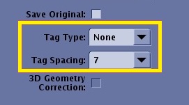
- VPS: As the Views per Segment increase, scan time and the number of cardiac phases decrease and edge blurring increases.
User CVs
Click the Advance tab to view the available User CVs. The CVs may vary based on the field strength and selected scan and imaging parameters.