• Object ID: 00000018WIA30D0E870GYZ
  • Topic ID: id_40025031 Version: 1.2
  • Date: Dec 21, 2021 6:42:23 PM

Patent foramen ovale

About this task

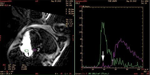
Real Time scans with the IR Prepared Imaging Option turned on can be used for detection of PFO. When the patient performs the valsalva maneuver, the blood flow shunt between the atria is elicited. If the realtime scan is acquired during the valsalva maneuver, the shunt can be imaged. IR-Prepared provides the necessary T1-weighted contrast. High temporal resolution is required with these scans because the shunt duration is typically less than one second. Achieve high temporal resolution by trading off high spatial resolution. A large FOV and slice thickness, small matrix values, and fractional NEX may be necessary to achieve the desired temporal resolution of four frames per second.

Blood clots traversing a PFO can be contributors to cerebral emboli, particularly with young patients. PFOs are difficult to identify because the transient amount of shunted blood is small and the shunt can only be visualized when the patient bears down using a valsalva maneuver. Therefore, this evaluation is performed using real time scanning.

Step-by-step instructions

  1. Explain the valsalva maneuver.
    • Carefully explain that the patient must bear down for 20 seconds and then release and begin breathing normally.
    • Since this maneuver must occur during real time scanning, which is very noisy, providing the patient with a hand signal to start and stop the maneuver is critical.
  2. Prescribe a Real Time acquisition.
    • TE = Min Full, Flip Angle = 30, Bandwidth = 31.25, FOV = 48, Slice Thickness = 20, Slice Spacing = 0, Frequency = 64, Phase = 64, NEX = 0.5, Phase FOV = 0.75, Shim = Auto
    • Click IR in the Contrast area of the Real Time Acquire tab.
  3. Acquire the Real Time scan with the valsalva maneuver.
    1. Use the real-time controls to define a 4-chamber view.
    2. Rotate the heart so that the apex is pointing up at the 12:00 position.
    3. Pause the real-time scan to communicate with the patient that the procedure is going to begin.
    4. The operator at the console must communicate with the person attending the patient when the valsalva maneuver must begin and end.
    5. Stop the scan when the maneuver is completed.
    6. Save the acquisition BEFORE the Real Time screen is closed. Failing to do so will result in losing the images acquired during real-time scanning.
  4. Display a time/intensity curve.
    1. Open/launch READY View.
    2. Press the up/down arrow keys to page through the images until you display an image with the right and left atrium.
    3. Place an ROI in the right atrium (1) and an ROI in the left atrium (2).
    4. Press the Spacebar after you deposit each ROI to generate the time/intensity curve.
      Figure 1. ROI and time/intensity curve
  5. Analyze the curve for a PFO.
    • A PFO is typically demonstrated as an additional peak on the time/intensity curve.