Use these steps to acquire a prescan and shimming technique to improve gated 2D FIESTA sequences that are prescribed in a single slice group. Cardiac FIESTA imaging often results in off-resonance artifacts when the center frequency is incorrectly obtained. These artifacts appear as inhomogenous areas within the blood pool and as ghosting across the image. They are more prominent in areas of fast or turbulent blood flow.
Step-by-step instructions
Position the patient orientation based on the selected coil.
Use the Phased Array Cardiac coil or other coils that produce optimum coverage and SNR.
Choose a 2D FIESTA pulse sequence from your Site or GE protocol list.
The Sequential Imaging Option is automatically selected to acquire one slice at a time and multiple cardiac phases. Therefore, cross-talk is not a problem.
The minimum TR is selected automatically. The calculated TR is the minimum TR attainable and is based on SAR restrictions. The TR may be adjusted through changes to any of the following parameters: frequency matrix, FOV, slice thickness, and flip angle. You achieve the best image quality at TR ≤ 4 ms. The bandwidth is typically ≥ 125 to keep the TR < 4.
The Phase matrix can be > Frequency value, which can help to avoid increasing the TR above the values mentioned previously.
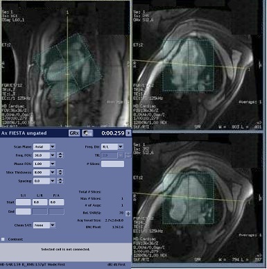
Place a small FOV shim volume over the heart.
The shim volume FOV should be approximately half the size of the scan FOV. For example, if the scan FOV is 36 cm, then the shim volume FOV should be 18 cm. For more details, see Shim volume procedure.
Figure 1. Shim volume over heart
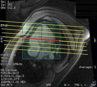
Place the parallel slices or single slice over the area of interest.
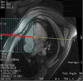
To optimize the gated 2D FIESTA prescan enhancement, the intersection of the shim volume and slice locations should only be placed over the anatomy of interest.
Figure 2. Correct intersection and placement of shim volume and slice locationsFigure 3. Incorrect intersection and placement of shim volume
Click Auto Prescan.
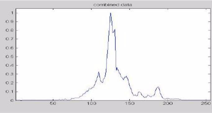
The enhanced gated 2D FIESTA prescan only applies to the intersection of the shim volume and the center slice of the graphic prescription. This ensures that the acquisition uses relevant frequencies centered on the area of interest.
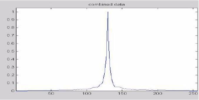
Frequencies outside the FOV that used to cause the spectrum degradation are thus eliminated with this technique. Figure 4. Center frequency spectrum for 2D FIESTAFigure 5. Center frequency spectrum for enhanced FIESTA sequence
Click Scan and view the results.
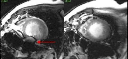
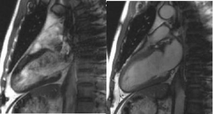
The image improvements are demonstrated in all gated 2D FIESTA scans. Figure 6. Reduced susceptibility artifact (right image)Figure 7. Off-resonance artifact at 3.0T (left) and reduced artifact (right)