- 00000018WIA30F56970GYZ
- id_400220221.2
- Jun 9, 2022 11:53:14 PM
Inhance: Deltaflow scan
Inhance Deltaflow is a non-contrast agent MRA method. It acquires two 3D slabs: one during systolic phase and one during diastolic phase. A multi-phase SSFSE scan is acquired to determine the diastolic trigger delay for the Inhance Deltaflow acquisition.
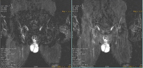
The signal produced from arterial flow is sensitive to the cardiac cycle. During systolic phase, arterial flow is fast resulting in a dark signal. During diastolic phase, arterial flow is significantly slower resulting in a bright signal. Unlike arterial flow, venous and background signal are relatively insensitive to the cardiac cycle. Subtraction of the systolic slab from the diastolic slab results in the visualization of the arteries with good background suppression. A STIR pulse can be applied to both the systolic and diastolic acquisition for additional fat suppression.
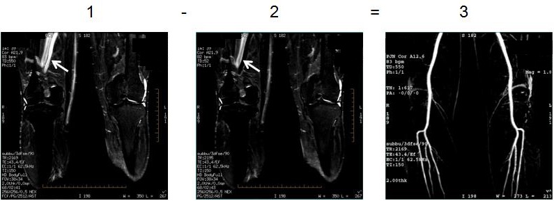
| Number | Description |
|---|---|
| 1 | Systolic |
| 2 | Diastolic |
| 3 | Subtracted image with artery visualization |
Use Inhance Deltaflow for peripheral run-off exams that do not use a contrast agent.
Consider this information when modifying Inhance Deltaflow scan parameters. For specific scan parameter values, select a protocol from your GE or Site library.
- Scan selections: 3D mode, Vascular family, Inhance DeltaFlow pulse.
General
- Inhance Deltaflow protocols are located in the GE library under Lower Extremities.
- With 3.0T systems, use a phased array coil to acquire signal from fast flowing iliac arteries. DeltaFlow is only suggested for the lower leg station at 3.0T. Other stations can result in image quality degradation due to B1 inhomogeneity effects. If you acquire a multi-station scan, be certain to carefully center each slab, evenly load the body coil, and use ASSET for the iliac stations. Given that the B1 inhomogeneity effects are very sensitive to anatomical body (weight and geometry), it may not work on every subject.
- It is strongly recommended that you review the unsubtracted source images to assess the vasculature and if needed, use other techniques to image the vessel of interest.
- At the end of the Inhance Deltaflow scan, a subtracted series is generated in the patient list: systolic images (first echo) are subtracted from diastolic images (second echo)). From the Session Apps list, select MRA to create projected images and reformatted axial images for further analysis.
- If you want to use the Pasting post processing option to paste images from multiple stations, only acquire coronal images. The Pasting algorithm does not support double-oblique images.
- The landmark location with 3.0T Inhance Deltaflow is extremely critical to the image quality. It is very important that the body coil is evenly loaded, particularly for the upper leg and pelvis stations. Improper landmarking results in signal lose due to coil loading issues.
Figure 2. Localizer for slab position for 3-stations 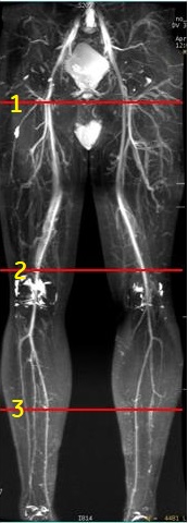
Table 2. Image legend Number Description 1 Center of the slab for station 1 2 Center of the slab for station 2 3 Center of the slab for station 3
Imaging Options
Flow Compensation: If the systolic flow is very slow, the subtracted image quality is compromised. Consider repeating the scan with Flow Compensation Imaging Option turned on. Flow Compensation uses higher crusher gradient settings in the frequency encoding direction for the systolic phase compared to the diastolic phase. a higher crusher setting produces flow spoiling in the systolic phase thereby reducing the arterial signal in the systolic phase. When the diastolic phase is subtracted from the systolic phase, the slow moving arteries are better visualized.Multi-phase SSFSE
Multiphase SSFSE acquires multiple phase images with increasing delay between each phase . An automatic subtraction of the first phase (corresponding to systolic) from other phase images provide arterial images, which can used to estimate the delay that corresponds to the optimum arterial visualization (diastolic start time).
Before you acquire the Inhance Deltaflow scan, acquire a multi-phase SSFSE scan to determine the optimum Diastolic Trigger Delay on the Cardiac tab. A multi-phase SSFSE series is included in the GE/Lower Extremities Inhance Deltaflow protocol. This scan is particularly important when the pathology occurs in one rather than both legs.
- Select either PG or ECG.
- From the Cardiac tab, select:
- RR intervals: 4 or 5 .
- Trigger Window: Auto or 20%. If the heart rate is very stable, use 10%.
- Trigger Delay: The Recommended Trigger Delay should be good for 90% of patients if the heart rate is less than 85 BPM. If the heart rate is fast (typically > 85 BPM), select a Trigger Delay from the multi-phase SSFSE scan. You can also enter your own Trigger Delay selected from the coronal SSFSE localizer.
- Inter-Seq Delay: typically select Even Space, which is incremental delay calculated by the number of phases and heart rate such that the temporal resolution is sampled within one RR interval. Minimum Delay is 80% of the Even Space.
- Diastolic Trigger Delay: View the multi-phase SSFSE images from the Viewer. Select the Trigger Delay based on the image that displays conspicuous arterial signal, the sharpest vessels, and 1-2 phases before any blurring/ghosting occurs. The Trigger Delay is displayed in the Patient List. Enter this value on the Cardiac tab. Use Recommended Delay if a multi-phase SSFSE scan was not acquired.
Figure 3. Multi-phase SSFSE image outline in yellow best demonstrates vessels of interest 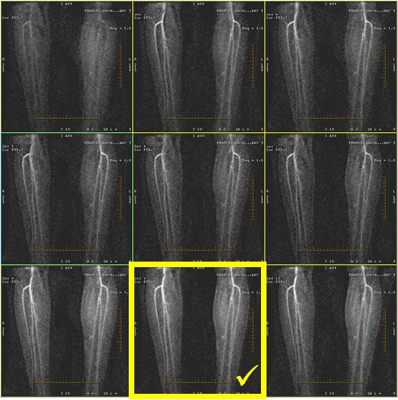
- Systolic Trigger Delay: typically select Recommended, which corresponds to Minimum with PG and 200 ms with ECG.
- If the patient's heart rate changes significantly between different stations, repeat the multi-phase SSFSE scan to determine the optimum diastolic trigger delay at each station.
- From the Multi-phase tab, select:
- Phases per Location: typically 15 is sufficient to visualize the vessels of interest
- Auto Subtract On
- 1st Phase of Same Series On
- Graphically prescribe a single coronal slice with the maximum allowed slice thickness (typically 150 mm) located over the vessels of interest.
Figure 4. Single coronal slice centered over vessel of interest 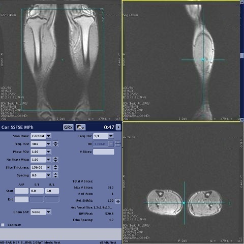
Deltaflow Graphic Rx
- Graphically prescribe a single coronal slab over anatomy of interest. Cover the entire anatomy to avoid phase wrap artifact.
Figure 5. Single coronal slab includes all anatomy of interest to avoid wrap artifact 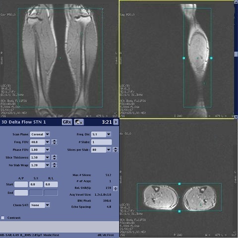
- For middle and upper stations, consider placing a posterior SAT pulse so that you can keep the slab size as small as possible for the optimum scan time. The SAT pulse is critical to reduce phase wrap when there is anatomy outside the FOV.
Figure 6. Exmaple of: Posterior SAT pulse positioned to minimize phase wrap 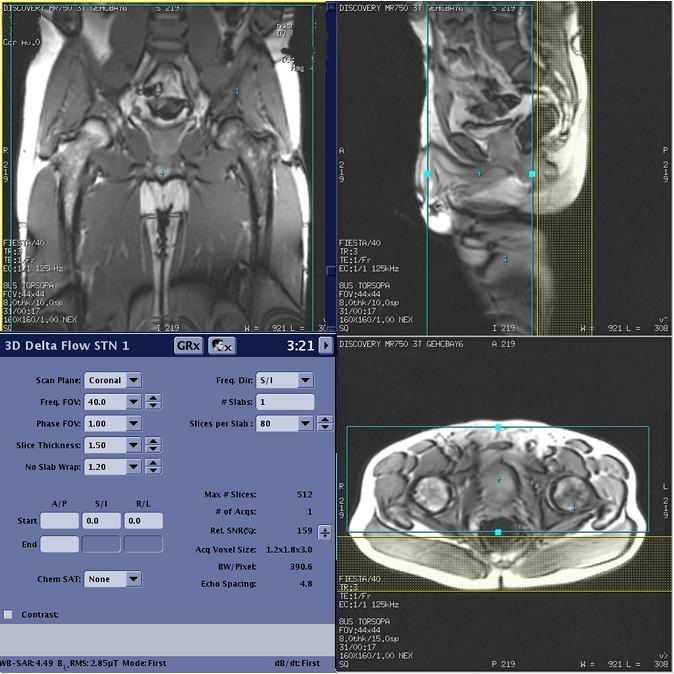
Figure 7. Left is pelvic image with no posterior SAT, right is image with posterior SAT pulse