

- 00000018WIA30815970GYZ
- id_400260011.2
- Aug 21, 2022 3:55:16 PM
Inversion Recovery scan
Inversion Recovery is a Spin Echo pulse sequence with an initial 180° inversion pulse prior to the 90° pulse. Use IR sequences produce T1-weighted or fat-suppressed images, particularly in abdomen or extremities. This sequence is also used for very heavily weighted T1 brain images.
The initial 180° pulse is used to create magnetization in the negative Z axis. The period between the 180° and 90° pulse is the inversion time (TI). The time between the 90° and the echo is TE, similar to a Spin Echo sequence.
- The effectiveness of the IR fat suppression technique varies based on changes in magnetic field homogeneity.
- Do not use IR pulse sequences with contrast agents because enhancing pathology could be suppressed if the shortened T1 effect corresponds to the null point.
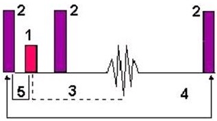
| Number | Description |
|---|---|
| 1 | 90° |
| 2 | 180° |
| 3 | TE |
| 4 | TR |
| 5 | TI |
The TI is the primary controller of image contrast. The TI value is determined by the time required for a tissue to recover from the negative longitudinal axis to the transverse plane. This time is the null point.
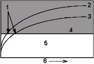
| Number | Description |
|---|---|
| 1 | Null |
| 2 | Fat curve |
| 3 | Water curve |
| 4 | Transverse plane |
| 5 | Contrast reversal zone |
| 6 | TI |
Use IR sequences to produce T1-weighted or fat-suppressed images, particularly in abdomen or extremities.
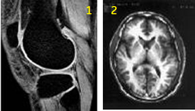
| Number | Description |
|---|---|
| 1 | IR knee image |
| 2 | IR brain image |
Consider this information when modifying Inversion Recovery scan parameters. For specific scan parameter values, select a protocol from your GE or Site library.
- Scan description: 2D Mode, IR pulse.