

- 00000018WIA30A38970GYZ
- id_400249191.3
- Mar 28, 2022 1:05:38 PM
ASL
ASL acquisition is a non-invasive, one-click application that allows whole brain CBF measurements. This contrast-free acquisition technique is ideal for patients in whom contrast in contraindicated.
The CBF maps are automatically displayed when a 3DASL series is selected and READY View is launched.
Algorithms
ASL READY View has algorithms that calculate Cerebral Blood Flow maps from a 3DASL scan.
Note the following:
- Water is a freely diffusible tracer.
- Homogeneous blood/brain partition coefficient for water is 0.9 ml/g.
- Labeling inversion efficiency is 80%.
The quantification algorithm is as follows:
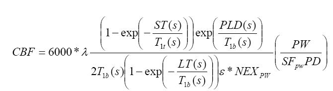
where, T1b is T1 of blood and is assumed to be 1.4s.
The partial saturation of the reference image (PD) is corrected for by using a T1t of 1.2s ( typical of gray matter). ST is saturation time and is set to 2s. The partition coefficient λ, is set to the whole brain average, 0.9. The efficiency, ε, is a combination of both inversion efficiency (0.8) and background suppression efficiency (0.75) resulting in an overall efficiency of 0.6. PLD is the post labeling delay used for the ASL experiment. LT is the labeling duration if it is set to 1.5s for the current version. PW is the perfusion weighted or the raw difference image. SFPW is the scaling factor of PW sequence. NEXPW is the number of excitation for PW images. The CBF is reported in ml/100gm/min units.
A threshold on the reference image (PD) is used to minimize the noise amplification in the resultant CBF images and is set as follows:
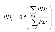
This allows us to look at the measure of the overall distribution of PD.
The default-scaling factor is 32. If the scaling factor for PW were other than 32, then it would be present in the PW header (0043,107F) 2nd value.
ASL measurement units
The ASL functional maps have the following units of measurement.
| Maps | Units |
|---|---|
| ASL-CBF | ml/100g/m |
READY View protocols that use ASL scan data
- ASL
- MR Brain