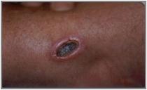
The following are a tips that will assist you in properly positioning and applying RF padding to your patients. Should you need more information on prevention of patient warming than what is provided here, refer to your surface coil and refer to Tissue Heating in this manual. If you need help beyond the documentation please do not hesitate to reach out to your local Applications Specialists.
Although the photos in the following sections are from a Discovery system, the safety padding guidelines apply to all MR systems.
Whole body padding
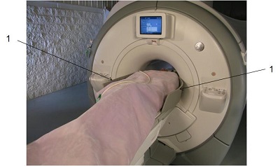
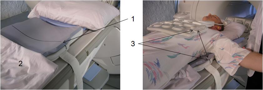
An important consideration when padding your patients is that you will need to double check the position of the pads once the patient is in the bore. Table movement may dislodge padding and expose skin to the scanner bore. Figure 2. Padding between patient and bore. 1 = bore pads
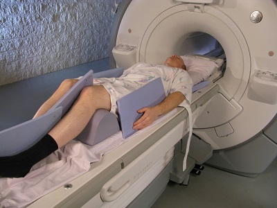
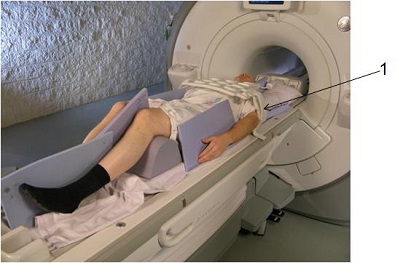
Notice that padding is positioned not only at the patient’s sides to prevent their arms from touching the bore, but that padding is also placed between the hands and thighs and between knees and ankles to prevent forming conductive loops. Figure 3. Patient padding
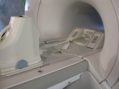
Surface coil padding
Padding with a surface coil presents different challenges from a patient RF padding perspective.
First rule of thumb is to remember to use all manufacturer provided padding to prevent motion and the patient’s skin from coming in contact with the coil, and to also use additional padding if appropriate to secure an opposing extremity to prevent contact with the coil which could also lead to burns or motion artifacts.
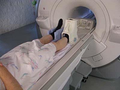
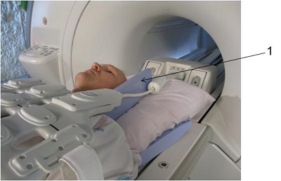
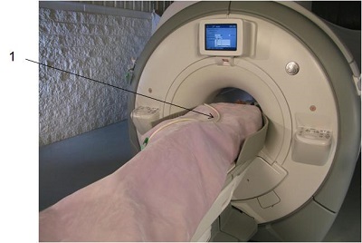
Just as with the whole body RF padding demonstration, you’ll need to make certain that the patient’s skin does not come into contact with the scanner bore and that padding is placed between the hands and thighs to prevent conductive loops. Figure 4. Extremity padding
A final safety consideration for surface coils is to ensure that the patient does not come into contact with the coil cable, therefore you may need to use additional RF padding to protect the patient.
Care should also be taken to ensure the cable is not looped in the bore and that it is routed down the center of the scanner bore. Figure 5. Coil cable with no loop
Cardiac coil padding
Follow your basic padding recommendations to prevent contact with the scanner bore and prevent conductive anatomical loops, but there are a couple of additional steps you’ll need to take to ensure patient safety
The cardiac coil does not require additional RF padding to be placed between the patient and the anterior coil component, but you should use the manufacturers pad on the posterior component of the coil for patient RF protection. You should also cover the patient with their gown before placing the anterior component of the coil and make certain both the anterior and posterior elements are in alignment. Figure 6. 1 = coil pad aligned with coil, 2 = sheet to cover pad, 3 = anterior and posterior coil elements aligned
Secure the coil snugly, but comfortably with the straps. Figure 7. Coil secured with straps
As is the case of all surface coils ensure that the cables do not come in contact with the patient and that they are not looped and routed down the center of the bore. As you can see there is significantly more cable that we need to isolate from the patient, so be sure to use as much padding as needed. Figure 8. 1 = Pad placed between cable and patient skin
If you are using the cardiac coil, it’s likely you are also using the ECG leads and cable. The rules for the ECG cable are the same as the coil cable. Route the ECG cable down the center of the bore, do not loop the ECG cable and do not allow it to come in contact with the coil cable. Figure 9. 1 = ECG cable with no loops