MAGiC artifacts may simulate pathology, which has the potential to lead to misinterpretation. Exercise caution when reviewing CSF spaces, its adjacent tissues and the posterior fossa, particularly for cases involving subtle pathology. MAGiC creates novel artifacts that are unique to synthetic images. Specifically, hyper-intensity or CSF suppression artifacts may be present on MAGiC T2 FLAIR. Hyper-intense signal may display as an artefactual edge enhancement between CSF and adjacent tissue. CSF suppression artifacts may appear as an unexpected bright signal. If in doubt, it is advisable to acquire a conventional 2D or 3D T2 FLAIR series or a MAGiC series in a different orientation for cross-sequence comparison. Presentation of traditional artifacts (such as motion) may not appear the same in conventional imaging due to signal sampling differences. For example, the appearance, frequency, and location of artifacts may be unconventional. Artifacts from patient movement, (e.g., due to sneezing, tremor, etc.) will propagate through all contrasts. Artifacts from physiological motion, such as arterial and CSF pulsations, ghosting, and any other interference during MAGiC acquisition, will impact all synthetic contrasts. Heightened attention to proper patient stabilization or immobilization is advised. Conventional troubleshooting via comparison to other sequences may not be appropriate.
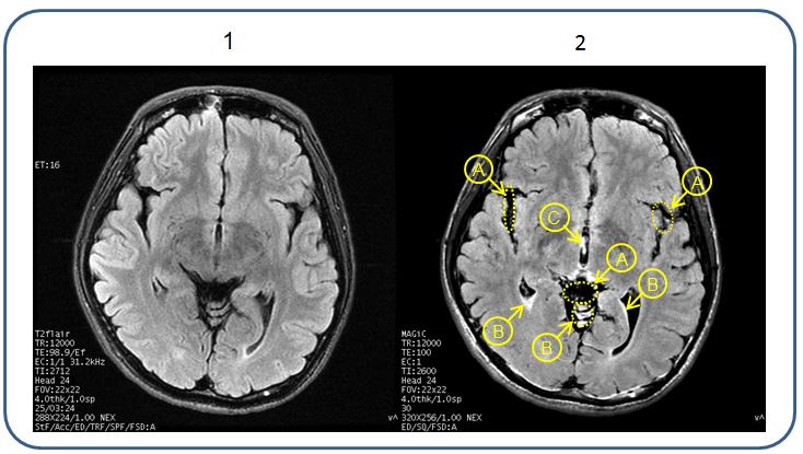
Figure 1 illustrates an example of differences between a MAGiC T2 FLAIR image compared with a conventional T2 FLAIR.
Figure 1. Conventional T2FLAIR image head versus a MAGiC T2W FLAIR image
Important: It may occur that a given MAGiC series does not display anatomy in exactly the same way as the conventional series. It is for this reason that the user is advised to consider all possible contrast weightings generated by MAGiC when performing image evaluation.
Table 1. Image legend
#
Description
1
Conventional T2 FLAIR
2
MAGiC T2W FLAIR
A = Hyperintense Vessel Sign (HVS)
B = High signal intensity at the edge of Cerebral Spinal Fluid (CSF)