The concern from time-varying gradient fields is to prevent cardiac stimulation and ventricular fibrillation. Cardiac stimulation in the most sensitive population percentile requires at least 39 times as much energy as is produced with peak gradient amplitude of 0.05 Tesla/meter and Slew Rate of 200 Tesla/meter/sec.
Regulatory bodies use avoidance of painful nerve stimulation to limit gradient output with an adequate safety margin. Painful nerve stimulation typically occurs at approximately double the mean stimulation perception threshold. Some discomfort is experienced about 1.5 times the mean PNS threshold, see figure below.
Peripheral nerve stimulation (PNS) problems are not of concern on systems compliant with IEC 60601-2-33 Normal or First Controlled Operating Modes. The IEC limits PNS to 80% of the mean threshold in Normal Mode (where stimulation should be rare) and 100% for the First Mode (where non-painful stimulation is expected in about half the patients).
If the patient can not tolerate PNS, change to Normal Mode to eliminate the problem. Otherwise change to a lower slew rate pulse sequence to continue scanning the patient. MR workers may experience similar sensations if they are in or very near gradient coils during active scanning, and so keep sufficient distance away from the gradient coils during scanning.
The Anterior/Posterior (A/P) (=Y) gradient axis typically has the lowest stimulation threshold. So it is prudent to keep the gradient waveform most likely to stimulate (the gradient waveform with the highest slew rate for the longest total ramp time) on a physical axis other than Y.
You should remain in constant contact with the patient, especially in the FIRST CONTROLLED MODE, to ensure that the patient does not feel painful stimulation (or high localized heating).
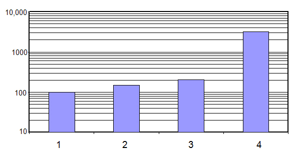
The figure below displays a graph of the relative mean threshold (vertical axis) and discomfort stimulation levels (horizontal axis) where relative means are for perception (100), discomfort (1000), and pain (10,000).
1 = threshold
2 = uncomfortable
3 = intolerable
4 = 1% cardiac
Figure 1. Relative mean threshold and discomfort stimulation levels
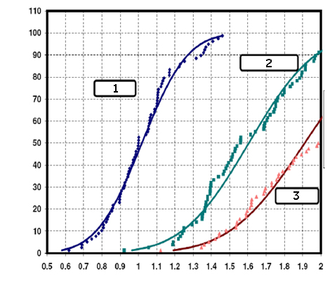
The distribution of those experiencing PNS is illustrated in the figure below; three curves where the horizontal axis is the normalized level and the vertical axis is the% probability of PNS. The curves represent the following:
1 = threshold
2 = uncomfortable
3 = intolerable
Figure 2. PNS probability. X axis = Fraction of the Mean Threshold (100% PNS). Y axis = Population percentile.
CAUTION
To reduce the possibility of PNS, make sure there is no skin–to–skin contact that can form a conductive loop through a part of the body, e.g.: inner thigh–to–thigh, calf–to–calf, hand–to–hand, hand–to–body, ankle–to–ankle contact.
CAUTION
Due to the rapid rate of change of the magnetic fields (dB/dt) used during some scans, a percentage of patients may experience a non-hazardous tingling or touch sensation. The PNS probability graph indicates the type of sensations caused at different percentages of the mean nerve stimulation threshold. Note that stimulation is relatively rare in NORMAL MODE (x-axis=0.8), but occurs about 50% of the time in the FIRST MODE (x axis=1). If this sensation is bothersome or uncomfortable to the patient, stop the scan. Change to NORMAL MODE to continue scanning the patient. The MR worker may experience similar sensations if remaining within the gradient field during active scanning. Exposure to gradient output can be minimized by keeping sufficient distance away (the distance required to complete necessary tasks in the scan room) from the gradient coils during scanning.
CAUTION
There is a possibility that mild peripheral nerve stimulation (PNS) may be induced in the MR worker when that person is exposed to the gradients when the system is operating in the First Level Controlled Operation Mode. The MR worker should remain outside the magnet room during scanning in this Mode except when circumstances dictate otherwise.
CAUTION
Peripheral nerve stimulation is not harmful. The potential for inducing peripheral nerve stimulation is kept within limitations. The MR system is limited from operating above 80% of the PNS threshold in the NORMAL Mode (100% of the mean PNS threshold in the First Mode) by the software (unless the system is in Second Controlled Mode). The point at which 50% of a population experiences PNS is the PNS threshold. PNS has been described as a light “touching” sensation felt on various areas of the skin surface. These areas vary depending upon which gradient axis is in use. Some common areas for the sensations are the bridge of the nose, arms, chest, and upper buttock/abdomen. Hands clasped together increase the potential for stimulation by approximately 65%. The potential for PNS is low, but it exists for all sequences in all gradient configurations.
Note: Please report all complaints of patient discomfort that may be associated with PNS during MR examinations (e.g., muscle twitches, tingling sensations, or headaches) to GE. See Incident reporting for contact information.