

- 00000018WIA30233970GYZ
- id_400261111.1
- Aug 6, 2021 9:32:28 AM
Restricted diffusion
The extra-cellular water in normal brain tissue diffuses freely, resulting in a dark signal. The extra-cellular water in dead brain tissue does not diffuse, resulting in a bright signal.
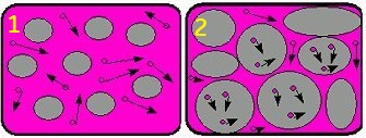
| # | Description |
|---|---|
| 1 | Normal diffusion |
| 2 | Restricted diffusion |
Use DW EPI for examining the tissues in the brain, liver, breast and prostate. The Diffusion tab only appears if the protocol has DWI EPI or DW EPI Tensor selected as the pulse sequence.
| Parameter | Description |
|---|---|
| b-values | The B-Value indicates the strength and sensitivity of motion probing gradients. As the B-value increases, the sensitivity of the motion probing gradients increases. The maximum B-value for DTI is 4000 s/mm2, the minimum B-value is 10 s/mm2. The text field allows # of b-values to be entered, which are displayed in the table. You can edit the values in the table. |
| NEX for T2 | This allows you to prescribe a unique NEX value for T2 and b-values. Typically use higher values (4-8). |
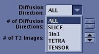
| Diffusion Direction
| For DTI imaging, you must choose TENSOR for the Diffusion Direction to allow the acquisition to be in tensor mode. Other direction options are not supported for DTI. For DWI, the Diffusion Direction allows you to define the direction of diffusion: All, L/R, A/P, or S/I for orthogonal planes; All or Slice for oblique planes. The diffusion images reflect the motion of water molecules in the selected diffusion direction. 3 in 1 acquires a single direction DWI with three diffusion gradients applied simultaneously. TETRA (tetrahedral) applies three axes simultaneously for each of the four diffusion directions. |
| # of Diffusion Directions | Enter the # of Diffusion Directions for the sequence. The maximum number of diffusion directions is 300, the minimum number is 6. The more directions prescribed, the higher your scan time. |
| # of T2 Images | Enter the # of T2 Images to be collected at the beginning of the acquisition. The maximum number that can be prescribed is 10, the minimum is 1. The recommended number of T2 images is 1. The more T2 images prescribed, the longer the scan time. |
| # of Synthetic b-values | Allows you to synthesize b-value diffusion weighted images using scanned DWI data. MAGiC DWI uses acquired DWI images to synthesize new DWI images with user selected b-values. |
| Recon All Images | Defaults to on and is not de-selectable. |
| Dual Spin Echo | Turns on an eddy current compensation technique, which reduces distortion. This option increases TE and decreases SNR. It only produces a single echo. For abdominal scans, typically de-select the Dual Spin Echo option, which produces the shortest TE that is needed for bowel gas considerations and also increases SNR. |
Consider this information when modifying DWI and DTI scan parameters. For specific scan parameter values, select a protocol from your GE or Site library.
- Scan selections: 2D Mode, Echo Planar Imaging family, DW EPI pulse.